Home
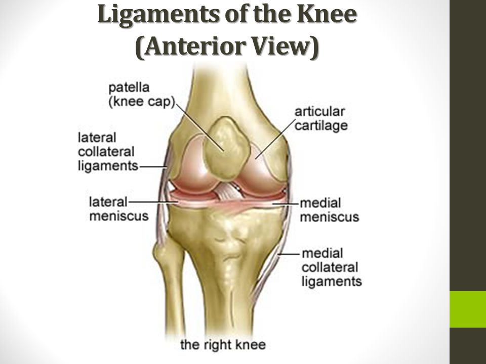
/ Anterior View Of Right Knee : Facebook : The patella (kneecap) sits over the front of the knee joint.
Anterior View Of Right Knee : Facebook : The patella (kneecap) sits over the front of the knee joint.
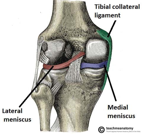
Anterior View Of Right Knee : Facebook : The patella (kneecap) sits over the front of the knee joint.. The medial and lateral meniscus come into view and the it band on the side. Pectoralis major lies anterior to pectoralis minor. The head is superior to the neck; Lateral meniscus anterior cruciate ligament posterior cruciate ligament patellar surface of femur transverse ligament tibial (medial) collateral ligament posterior cruciate ligament lateral condyle lateral meniscus medial. Learn vocabulary, terms, and more with flashcards, games, and other study tools.
Extending along the anterior surface of the thigh are the four. Natural color photograph of dissection of the right knee, anterior view: For more anatomy content please follow us and visit our website: Anatomynote.com found knee anatomy in detail anterior view from plenty of anatomical pictures on the internet. An acl tear often leads to the knee giving out, and may require surgical.
Anatomy Of The Knee Ppt Download from slideplayer.com For more anatomy content please follow us and visit our website: The anterior cruciate ligament (acl) is one of the key ligaments that help stabilize your knee joint. We hope this picture knee anatomy in detail anterior view can help you study and research. The head is superior to the neck; Anatomynote.com found knee anatomy in detail anterior view from plenty of anatomical pictures on the internet. Four major ligaments connect the bones and stabilize the knee joint. The patella (kneecap) sits over the front of the knee joint. As the image moves deeper you can begin to see the medial collateral ligament along the tibia and the anterior cruciate ligament.
The femur (thigh bone) contacts the tibia (shin bone) at the knee joint.
It's most commonly torn during sports that involve sudden stops and changes in direction — such as basketball, soccer, tennis and volleyball. For more anatomy content please follow us and. This is an online quiz called anterior view of right knee. The knee is stabilized by a pair of cruciate ligaments. Natural color photograph of dissection of the right knee, anterior view: Normal human anatomy of a knee, front view. There is a printable worksheet available for download here so you can take the quiz with pen and paper. Learn vocabulary, terms, and more with flashcards, games, and other study tools. Extending along the anterior surface of the thigh are the four. Just need a glimpse, leave your valuable advice let us know , and subscribe us! There is a printable worksheet available for download here so you can take the quiz with pen and paper. The acl is responsible for a large part of the knee's stability. Four major ligaments connect the bones and stabilize the knee joint.
Click on the tags below to find other quizzes on the same subject. Learn vocabulary, terms, and more with flashcards, games, and other study tools. These motions of the knee allow the body to perform such important movements as walking, running, kicking, and jumping. Normal human anatomy of a knee, front view. As the image moves deeper you can begin to see the medial collateral ligament along the tibia and the anterior cruciate ligament.
The Knee Joint Articulations Movements Injuries Teachmeanatomy from teachmeanatomy.info The acl is critically important because it prevents the tibia from being pushed too far anterior relative to the femur. Giant cell tumor of the right tibial. Anterior view of the right knee. The knee is stabilized by a pair of cruciate ligaments. The acl plays an important role in preventing hyperextension of the knee by limiting the anterior movement of the tibia. For more anatomy content please follow us and. Extending along the anterior surface of the thigh are the four. The anterior cruciate ligament (acl) stretches from the lateral condyle of femur to the anterior intercondylar area.
These motions of the knee allow the body to perform such important movements as walking, running, kicking, and jumping.
The anterior cruciate ligament (acl) is one of the key ligaments that help stabilize your knee joint. Giant cell tumor of the right tibial. The medial and lateral meniscus come into view and the it band on the side. Damage in even one part can hinder the functioning of the knee. The patella is located anteriorly in the lower limb. Learn vocabulary, terms, and more with flashcards, games, and other study tools. Learn vocabulary, terms, and more with flashcards, games, and other study tools. Anterior view of flexed right knee fibular collateral ligament anterior cruciate ligament patellar ligament anular ligament lateral meniscus posterior cruciate ligament tibiofibular ligament medial meniscus lliofemoral ligament tibial collateral ligament fibula tibia zoom reset. Click on the tags below to find other quizzes on the same subject. We are pleased to provide you with the picture named knee anatomy anterior view and lateral view in detail.we hope this picture knee anatomy anterior view and lateral view in detail can help you study and research. You may also find tibia, medial meniscus, medial collateral ligament, articular surface of the tibia, a posterior cruciate ligament as well. Anterior deep view of right knee joint study guide by ryanflora5 includes 14 questions covering vocabulary, terms and more. Lateral meniscus anterior cruciate ligament posterior cruciate ligament patellar surface of femur transverse ligament tibial (medial) collateral ligament posterior cruciate ligament lateral condyle lateral meniscus medial.
Right knee anatomy anterior view in this image, you will find medial condyle, articular cartilage, posterior cruciate ligament, medial meniscus, medial collateral ligaments, tibia in it. Knee joint is one of the most important hinge joints of our body. The muscles of the knee include the quadriceps, hamstrings, and the muscles of the calf. Its complexity and its efficiency is the best example of god's creation. 7.84 anterior/lateral view of right leg and foot anterior dorsiflex the ankle (talocrural joint) invert the foot middle, anterior surface of fibula and interosseous membrane distal phalanx of first toe deep peroneal 1_4, 5, sl extensor digitorum longus extensor hallucis longus extensor hallucis longus extensor digitorum longus
Knee Sprains And Meniscal Injuries Injuries Poisoning Msd Manual Professional Edition from www.msdmanuals.com This is an online quiz called anterior view of the right knee. Anatomy of the knee joint (sagittal view) in addition, the knee joint is strengthened by various ligaments, such as the patellar ligament, tibial and fibular collateral ligaments, and oblique popliteal ligament. You will also find patella, trochlea, posterior cruciate ligament, medial collateral ligament, medial meniscus, tibial plateau, tibia, tibial. In this image, you will find femur, anterior cruciate ligament, lateral femoral condyle, lateral meniscus, lateral collateral ligament, fibula in it. Click on the tags below to find other quizzes on the same subject. An acl tear often leads to the knee giving out, and may require surgical. The acl becomes more clear as you more deeper into the knee. Just need a glimpse, leave your valuable advice let us know , and subscribe us!
Rehman, irving smith, chadwick f.
7.84 anterior/lateral view of right leg and foot anterior dorsiflex the ankle (talocrural joint) invert the foot middle, anterior surface of fibula and interosseous membrane distal phalanx of first toe deep peroneal 1_4, 5, sl extensor digitorum longus extensor hallucis longus extensor hallucis longus extensor digitorum longus In the image above, the physician is pointing to the anterior cruciate ligament, or acl, one of these important ligaments. An acl tear often leads to the knee giving out, and may require surgical. The head is superior to the neck; See more ideas about anatomy, knee, knee pain. Anatomynote.com found knee anatomy in detail anterior view from plenty of anatomical pictures on the internet. Giant cell tumor of the right tibial. Inside the joint, there are additional reinforcing ligaments, such as the transverse ligament, together with the anterior (acl) and. Transcribed image textfrom this question. We think this is the most useful anatomy picture that you. You may also find tibia, medial meniscus, medial collateral ligament, articular surface of the tibia, a posterior cruciate ligament as well. For more anatomy content please follow us and. Our latest youtube film is ready to run.
These motions of the knee allow the body to perform such important movements as walking, running, kicking, and jumping anterior view of knee. The acl plays an important role in preventing hyperextension of the knee by limiting the anterior movement of the tibia.